“ MRI findings in patients with suspected medial meniscus tears were inaccurate in 25% of cases and patients with suspected anterior cruciate ligament tears were inaccurate in 22% of cases, after arthroscopy revealed no tears."
- Smith, Christian, et al.
Understanding the Limitations of MRI
Magnetic Resonance Imaging (MRI) has revolutionized the field of medical diagnostics, providing detailed images of the human body without the use of radiation. While it has become an invaluable tool in healthcare, we want to consider that like any technology, MRI has its limitations. Understanding these limitations is crucial for both healthcare professionals and patients alike to help prevent inaccurate diagnosis and unnecessary interventions.
Here is one example to set the stage. Just this past week I was checking in on a past patient and she is a college athlete and so she was telling me about a recent injury. She had an MRI and the doctor said she would need surgery. Naturally, this can be life changing news for that athlete! She may not have had all these thoughts but it is not farfetched for her to wonder if she will ever be able to play again? Will she keep her scholarship? If not, will she stay at that school and then who knows how that might change a life trajectory. So we see how these opinions can carry weight. Well, she wisely went and got a second opinion and to her surprise, that physician said he does not think she needs surgery! So, what's going on here?! Well it's not often talked about how many variables can affect the final recommendation with an MRI. So today we will dig into that a little to give you a more balanced perspective. But what this second opinion provided for that athlete is freedom. Freedom to now make a decision that she decides is best for her with the knowledge that both routes could be acceptable routes.
-
Interpretation Error: A clear image is not always possible but even when it is clear, there still can be human error in interpreting the picture (1). The study by Herzog et al. had a single person with back pain go to 10 different MRI locations and they found that the results were highly variable based on the facility and radiologist who read the results. That individual received 49 distinct findings. No single finding existed across all locations and only one finding was reported on 9/10 reports.
-
Positioning Limitations: Another problem is most MRIs are taken lying down. Ligament, tendon and muscle structures will change drastically with standing or when moving (2). Weight Bearing and Dynamic-Kinetic MRI exist but are rarely used due to availability, feasibility and expense.
-
Metallic Interference: One of the most well-known limitations of MRI is its sensitivity to metal. The strong magnetic fields generated during an MRI can interact with metallic objects, leading to distortions in the images or, in extreme cases, causing harm to the patient. Patients with certain implants, such as pacemakers or metal fragments, may be advised against undergoing an MRI. Other motion artifacts or obesity can also affect the quality and clarity (4)
-
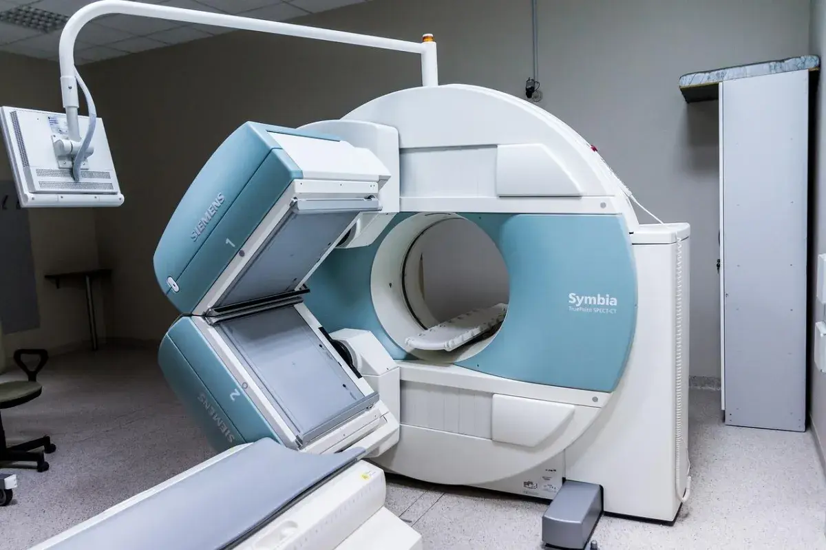
Claustrophobia and Patient Cooperation: MRI machines are often large, tube-like structures that can induce feelings of claustrophobia in some individuals. This can result in patient discomfort and difficulty remaining still during the procedure, impacting the quality of the images produced.
-
Functional Limitations: While MRI excels at providing detailed structural images, it has limitations in assessing certain functional aspects of the body. Functional MRI (fMRI) attempts to address this by measuring changes in blood flow, but it may not capture dynamic processes as effectively as other imaging techniques.
-
Expense and Availability: MRI technology is expensive to install and maintain, making it less accessible in some regions or healthcare settings. The cost of an MRI scan can be a barrier for some patients, and long wait times for appointments are not uncommon in areas with limited resources.
-
Contrast Agents: Contrast agents are sometimes used to enhance the visibility of certain structures in MRI images. However, some patients may be allergic to these contrast agents and they can also be toxic to the body, limiting their use.
While MRI is a powerful diagnostic tool, it's essential to recognize its limitations. Understanding these limitations allows patients and healthcare providers to make informed decisions about the most appropriate next step for each patient's unique situation.
For most musculoskeletal issues, we are able to gain the information we need from a physical exam. During the exam we can move through and assess the painful positions, we can test strength, mobility, and function of particular muscles. Then once we finish the exam we don't just hand you the findings but an action plan to get you back on your feet. Next time you are thinking of an MRI, consider a physical therapy evaluation with us and maybe we can save the expense, time and other risks associated.
References
-
Garland, L. Henry. “On the scientific evaluation of diagnostic procedures: Presidential address thirty-fourth annual meeting of the Radiological Society of North America.” Radiology 52.3 (1949): 309-328.
-
Herzog, Richard, et al. “Variability in diagnostic error rates of 10 MRI centers performing lumbar spine MRI examinations on the same patient within a 3-week period.” The Spine Journal17.4 (2017): 554-561.
-
Jinkins, J. Randy, Jay S. Dworkin, and Raymond V. Damadian. “Upright, weight-bearing, dynamic–kinetic MRI of the spine: initial results.” European radiology 15 (2005): 1815-1825.
-
Singh, Dinesh R., Michael SM Chin, and Wilfred CG Peh. “Artifacts in musculoskeletal MR imaging.” Seminars in musculoskeletal radiology. Vol. 18. No. 01. Thieme Medical Publishers, 2014.
-
Smith, Christian, et al. “Diagnostic efficacy of 3-T MRI for knee injuries using arthroscopy as a reference standard: a meta-analysis.” American Journal of Roentgenology 207.2 (2016): 369-377.


